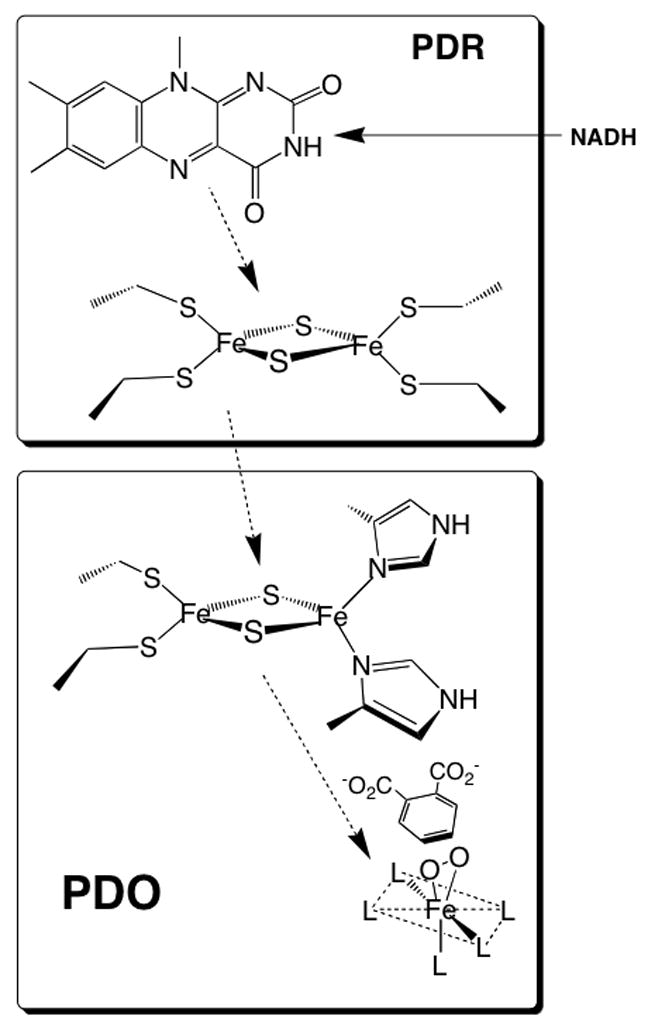
Figure 1.

Phthalate dioxygenase system. This figure shows the various redox cofactors and the general pathway for electron transfer. The ligands of the iron mononuclear center are represented by “L”. The figure also shows the substrate (phthalate) and an oxygen molecule bound to the iron side-on, a binding arrangement demonstrated for the NDO system [25]. Because no X-ray structure of PDO is available, assignment of ligands and geometry is not specified. However, the X-ray structures of related dioxygenases (see below) shows that the mononuclear site can be described as a 2-His 1-carboxylate “facial triad” [26]. Likely ligands in PDO are the well conserved His-181, His-186, and Asp-358. Other studies have indicated that one or two waters are also ligated in various states of PDO[27].
