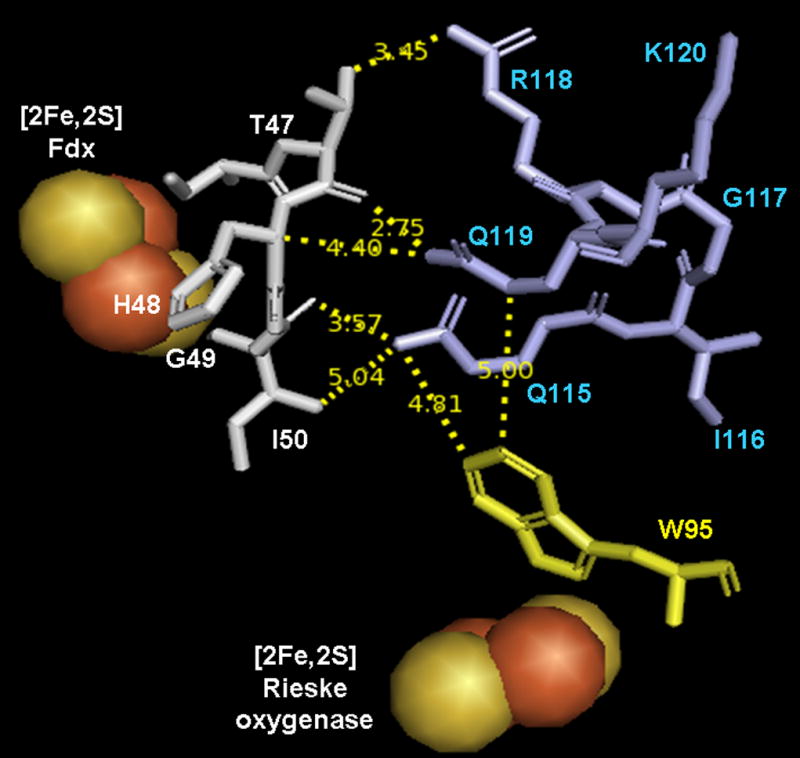
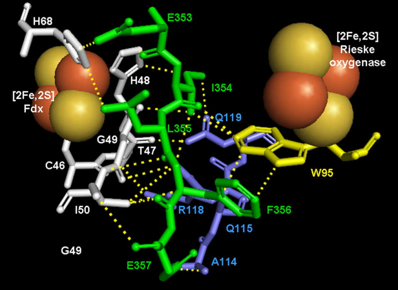
Figure 3.


Amino acid arrangement in the vicinity of the [2Fe-2S] Rieske center of CarDO in its complex with Fdx (based on 2DE5). Fdx residues are shown in white, residues of CarDO subunits are shown in light blue if belonging to the same monomer as the Rieske center shown and in green if belonging to an adjacent oxygenase subunit. Conserved Trp 95 (Trp94 in PDO) is shown in yellow. A and B represent different views of the amino acids arrangement with A highlighting the interaction of Trp95 with the same subunit residues and B highlighting Trp95 interactions with residues from the adjacent oxygenase subunit. Dotted lines on B represent short range interactions (<4 Å) between selected residues. Figures were built in PyMOL 2006 from DeLano Scientific Inc., which incorporated Open-Source PyMOL 099rc6.
