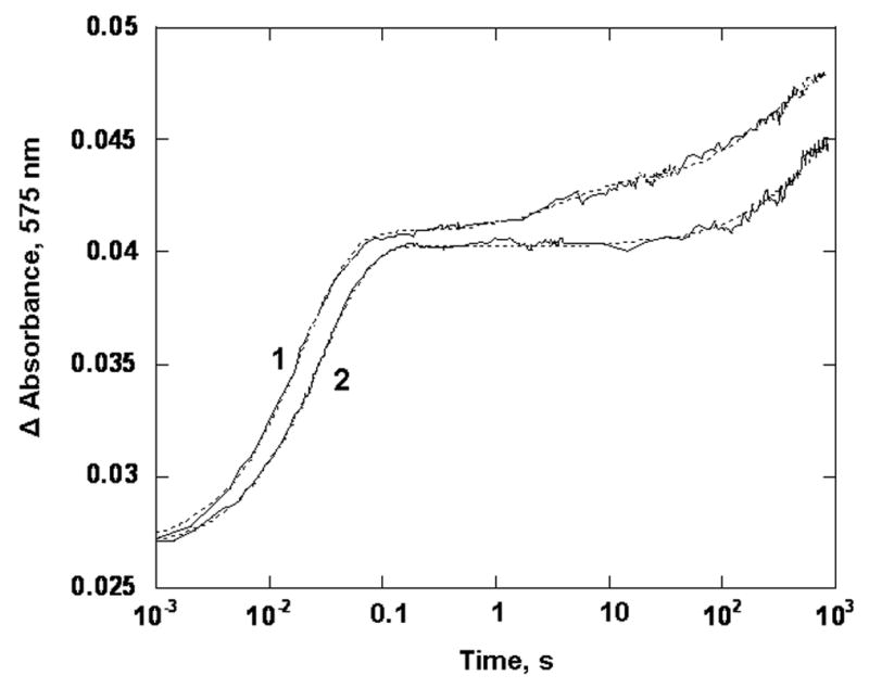
Figure 5.

Oxidation of reduced PDO by oxygen. PDO (20 μM) was mixed with 250 μM O2 in 0.1M HEPES pH 7.8, in the presence of 3 mM phthalate at 22 °C. Presented are experimental trances (solid line) and the fits obtained with the parallel reaction model (dotted lines) for WT PDO (1) and the W94Y variant (2), All concentrations are those before mixing in the stopped-flow spectrophotometer. Traces were recorded at 575 nm and truncated at 900 s for presentation.
