
Figure 1. Effect of Adenoviruses on HMVEC-L growth.
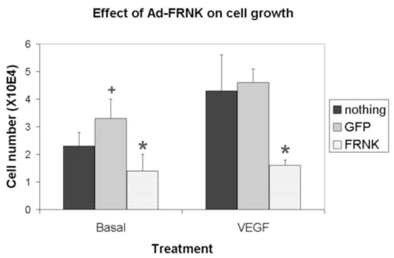
Cells (passage 2–3) were trypsinized, replated into 12-well plates (2 × 10⁴ cells per well), and incubated overnight. The cells were washed and the medium was replaced with serum-free medium, which contained nothing, Ad-GFP (5 pfu/cell), or Ad-GFP-FRNK (5 pfu/cell). The cells were then incubated overnight. After virus infection was confirmed using a fluorescent microscope VEGF (100 ng/ml) was added to some wells. After 72 hours, the cells were trypsinzed and manually counted using a hemotocytometer. Shown is mean ± standard error for triplicate wells of a representative experiment.
*, significantly different than Ad-GFP; +, significantly different than no virus