Fig. 1.
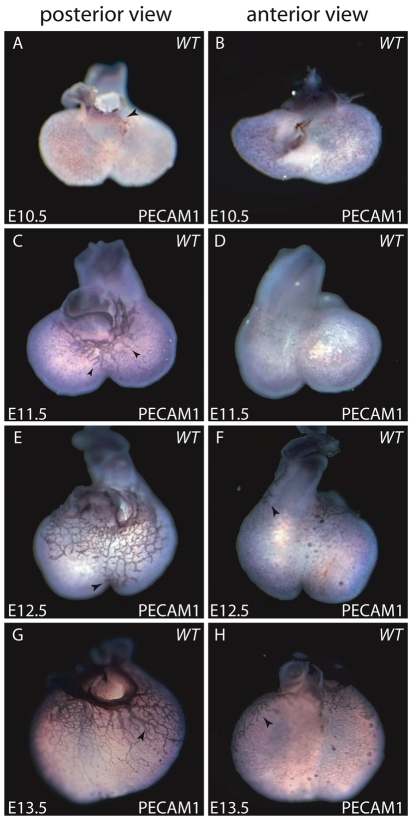
Vascular patterning of the coronaries during mouse embryonic development. (A,B) PECAM1-positive cells (purple) observed on the posterior (A) and anterior (B) surface of the heart by E10.5. Some endothelial cells are present at the atrioventricular junction (arrowhead in A). (C,D) PECAM1 staining showing nascent coronary vessels on the posterior (C) and anterior (D) surface of the heart at E11.5. Arrowheads in C indicate the distal end of the endothelial tubes extending from the atrioventricular junction to the apical region of the heart. (E,F) PECAM1 staining of the endothelial network on the posterior (E) and anterior (F) surface of E12.5 embryonic hearts. By E12.5, the vascular endothelial network has reached more apically to the interventricular region of the heart (arrowhead in E). Also, some vessels appear laterally and begin to extend to the anterior surface of the heart (arrowhead in F). (G,H) PECAM1 staining showing the pattern of coronary endothelial cells on the posterior (G) and anterior (H) surface of the heart at E13.5. More mature coronary vessels are formed on the posterior surface by E13.5 (arrowhead in G), and some vessels have extended to the anterolateral surface of the heart (arrowhead in H).

