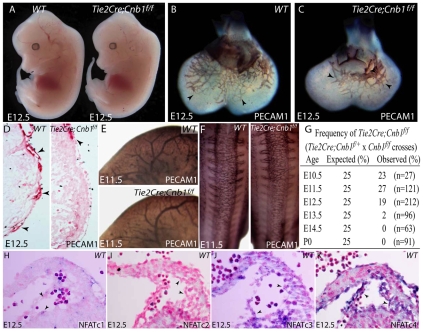
Fig. 4.
Endothelial calcineurin-NFAT signaling is required for normal coronary vessel patterning. (A) Gross morphology of wild-type (left) and Tie2-Cre;Cnb1f/f (right) embryos at E12.5. (B,C) PECAM1 whole-mount staining of endothelial cells in wild-type (B) and Tie2-Cre;Cnb1f/f (C) hearts at E12.5. Arrowheads denote endothelial cells. (D) Sections of wild-type (left) and Tie2-Cre;Cnb1f/f (right) hearts showing distribution of nascent coronary vessels (arrowheads). Red, PECAM1 staining; pink, counterstaining with nuclear Fast Red. (E) PECAM1 staining of E11.5 wild-type (upper panel) and Tie2-Cre;Cnb1f/f (lower panel) embryos showing normal vasculature in the cranial region. (F) PECAM1 staining of E11.5 wild-type (left) and Tie2-Cre;Cnb1f/f (right) embryos showing normal vasculature in the dorsal region. (G) Frequency of Tie2-Cre;Cnb1f/f embryos harvested at different embryonic dates compared with the expected mendelian ratio. (H-K) RNA in situ hybridization for NFATc1 (H), c2 (I), c3 (J) and c4 (K) transcripts (blue) on coronary vessels of E12.5 wild-type embryos. Arrowheads point to coronary endothelial cells. Pink, counterstaining with nuclear Fast Red.

