Fig. 2.
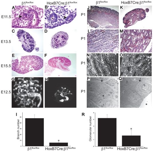
HoxB7Cre;β1flox/flox mice have a severe branching morphogenesis phenotype. (A,B) Decreased branching of the UB is present in E11.5 HoxB7Cre;β1flox/flox embryos (600×). The Wolffian duct is shown with an arrowhead and the UB by an asterisk. (C-F) Decreased UB branching and nephron number is present in E13.5 (C,D) and E15.5 (E,F) HoxB7Cre;β1flox/flox kidneys when compared with β1flox/flox kidneys (40×). (G-I) Cultures of E12.5 kidneys of HoxB7Cre;β1flox/flox and β1flox/flox mice were performed on transwells, as described in the Materials and methods. The kidneys were stained with antibodies directed against E-cadherin (G,H). The number of branches was counted in 10 kidneys from both genotypes and expressed as mean ± s.d. Differences between HoxB7Cre;β1flox/flox and β1flox/flox mice (*) were significant with P<0.01 (I). (J,K) PAS staining revealed that P1 HoxB7Cre;β1flox/flox mice have small kidneys, fewer nephrons and dilated tubules in both the cortex and medulla when compared with β1flox/flox mice (25×). (L,M) Collecting ducts within the papilla of P1 HoxB7Cre;β1flox/flox mice were dilated and irregular (200×). (N,O) E-cadherin staining of P1 kidneys showed no differences in localization and cell polarity between HoxB7Cre;β1flox/flox and β1flox/flox mice. (P,Q) Electron microscopy of collecting ducts demonstrated dilatation of the tubules in P1 HoxB7Cre;β1flox/flox mice, but there were no abnormalities in epithelial cell polarization or basement membrane structure (8500×). The arrows mark the tubular basement membrane and the asterisks demonstrate the tubular lumen. (R) The number of glomeruli in the cortices from similar sections of 10 P1 HoxB7Cre;β1flox/flox and β1flox/flox mice were counted and expressed as glomeruli/mm2. The mean and ±s.d. are shown. Differences between HoxB7Cre;β1flox/flox and β1flox/flox mice (*) were significant, with P<0.01.

