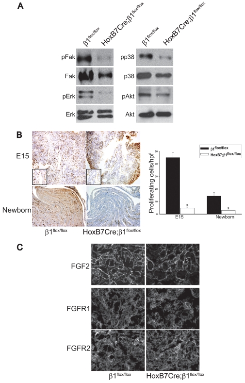
Fig. 6.
HoxB7Cre;β1flox/flox mice have severe signaling and proliferative defects. (A) Medullas of P1 β1flox/flox and HoxB7Cre;β1flox/flox kidneys were lysed and 20 μg of total protein was analyzed by western blot for levels of activated and total FAK, ERK, p38 MAPK and Akt. (B) Ki67 staining was performed on kidneys of β1flox/flox and HoxB7Cre;β1flox/flox E15.5 and newborn mice. The number of Ki67-positive cells in the UB (E15.5) or collecting ducts (newborn) of the mice was quantified and expressed as mean ± s.d. of five high power fields of three different mice. Asterisks indicate statistically significant differences (P<0.05) between HoxB7Cre;β1flox/flox and β1flox/flox mice. (C) Tubules derived from both the metanephric mesenchyme and ureteric bud of E15.5 β1flox/flox and HoxB7Cre;β1flox/flox kidneys were immunostained for FGF2, FGFR1 and FGFR2.

