Figure 2.
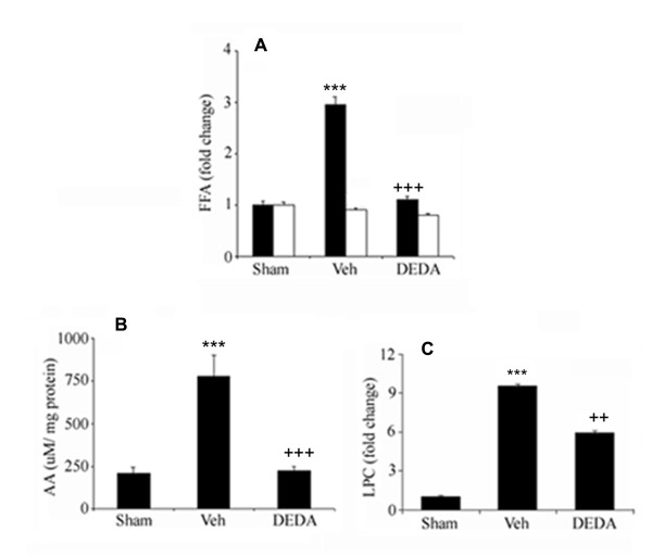
Effect of IR injury and DEDA treatment on the levels of FFA, AA and LPC in brain tissue at 24 hours of reperfusion after 60 min MCAO. Levels of FFA (A) and AA (B) were measured using GC and content of LPC (C) was quantitated by HPTLC. Data are expressed as means ± SD from triplicate determinations of 5 different samples (n = 5). Closed bars represent ipsilateral and open bars represent contralateral hemispheres. *** p < 0.001 vs. Sham, +++ p < 0.001 vs. Veh and ++ p < 0.01 vs. Veh.

