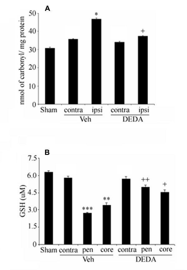
Figure 4.
Effect of IR and DEDA on levels of PC and GSH in brain tissue at 24 hours of reperfusion after 60 min MCAO. Levels of protein carbonyl were significantly elevated in the ipsilateral from the Veh brain. Treatment with DEDA reduced the PC level (A). IR significantly depleted the level of GSH in the penumbra and treatment with DEDA attenuated the GSH content in the Veh brain (B). Data are expressed as means ± SD from triplicate determinations of four different samples in each group. *p < 0.05 vs. Sham, *** p < 0.001 and ** p < 0.01 vs. Sham, ++ p < 0.01 and + p < 0.05 vs. Veh.

