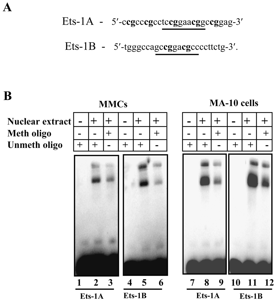
Fig. 8. Effect of methylation on in vitro Ets-1 binding in Npr1 promoter.
A) Schematic diagram showing the sequence of the wild-type Ets-1A and Ets-1B oligonucleotides used for EMSA. Sequences in bold show the CpG sites recognized by SssI methylase. Underlined sequences show the Ets-1A and Ets-1B consensus sites. B) The gel retardation assay in nuclear extract from MMCs and MA-10 cells using unmethylated and methylated Ets-1A and Ets-1B oligonucleotides. Lanes 1, 4, 7, and 10 contain free probe. Lanes 2 and 5 show MMCs nuclear protein complex binding with Ets-1A and Ets-1B sites, respectively, and lanes 8 and 11 show the MA-10 cells nuclear protein complex binding with Ets-1A and Ets-1B sites, respectively. Lanes 3 and 6 show the binding reaction of MMCs nuclear protein extract with methylated Ets-1A and Ets-1B probes, respectively, and lanes 9 and 12 show binding reactions with MA-10 nuclear protein. Meth oligo indicates methylated oligonucleotide and Unmeth oligo indicates unmethylated oligonucleotides.

