
Figure 9. Trisk 49 and Trisk 32 expression during differentiation of myoblasts.
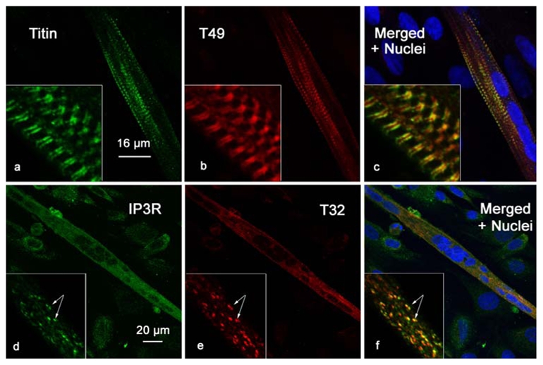
Primary cultures of satellite cells were induced in differentiation. When myotubes formed, the cells were fixed and labeled with antibodies against titin and Trisk 49 (panels a, b and c), or IP3R and Trisk 32 (panels d, e and f). The nuclei were stained with TO-PRO 3 (blue labeling in panels c and f). Titin and Trisk 49 antibodies labeled only myotubes (aligned nuclei) and not myoblasts (isolated nuclei), and both presented a striated pattern. IP3R and Trisk 32 antibodies label the myotubes (multinucleated cells) and the myoblasts (isolated nuclei), with a diffuse punctuated pattern.