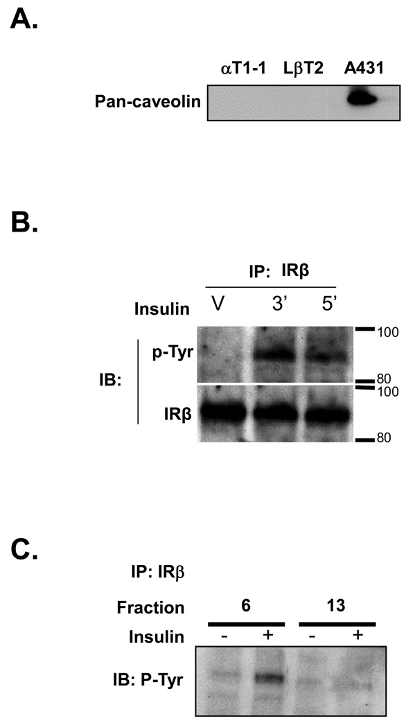
Figure 2. Insulin receptor is phosphorylated in a non-caveolar lipid raft compartment.
(A.) Whole cell RIPA lysates were prepared from either αT1-1, LβT2 or A431 cells. Western blots were conducted using a pan-caveolin antibody that detects caveolin-1, caveolin-2 and caveolin-3. (B.) Western Blot analysis of IRβ immunoprecipitated from LβT2 cell protein extracts. Cells were treated with vehicle (V) for 5 min or insulin for 3 or 5 min and then harvested. Extracts were immunoprecipitated with anti-IRβ and subjected to SDS-PAGE and Western Blotting with anti phospho-tyrosine (α-p-Tyr) antibody. The presence of IRβ was confirmed by stripping the membrane and re-blotting with anti-IRβ (IRβ). (C.) Western blot analysis of IRβ immunoprecipitated from LβT2 lipid raft fractions treated for 5 min with either vehicle or 10 nM insulin. Fraction 6 represents low-density (raft associated) proteins and fraction 13 represents high-density (non-raft associated) proteins.

