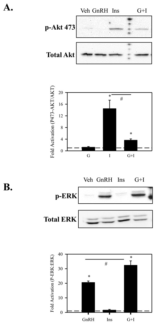
Figure 4. GnRH attenuates insulin induced Akt activation while insulin accentuates GnRH induced ERK activation.
(A.) LβT2 cells were serum starved for 4 h followed by treatment with either vehicle, 10nM GnRH (GnRH), 10 nM insulin (Ins), or both (G+I) for 15 min. Cells were then harvested in RIPA buffer and subjected to SDS-PAGE and western blotted using an anti-phosho-Akt (473) antibody. After probing with anti-p-Akt, blots were stripped and re-probed with an antibody that detects Akt independent of phosphorylation. (B.) LβT2 cells were serum starved for 4 h followed by treatment with either vehicle, 10nM GnRH (GnRH), 10nM insulin (Ins), or both (G+I) for 15 min. Cells were then harvested in RIPA buffer and subjected to SDS-PAGE and western blotted using an anti-phospho-ERK1/2 antibody. After probing with anti-p-ERK, blots were stripped and re-probed with an antibody that detects ERK 1/2 independent of phosphorylation.

