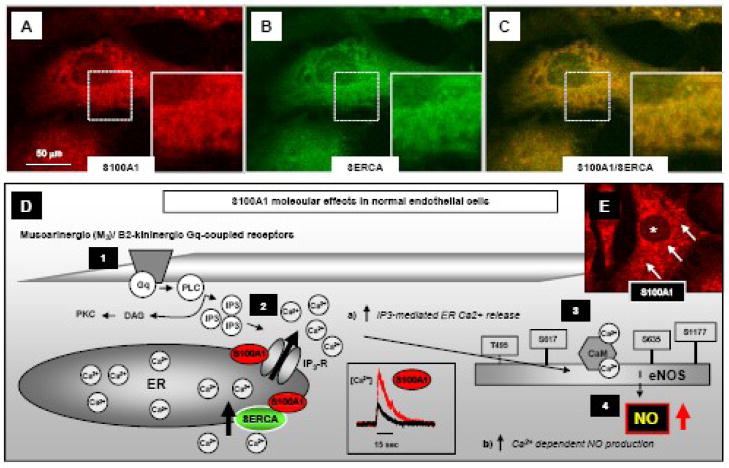
Figure 5. Subcellular S100A1 location and mechanisms conveying S100A1-mediated NO production in endothelial cells.
A–C, representative confocal fluorescent images highlight partial co-localization (yellow) of S100A1 (red) with SERCA (green) in an aortic mouse EC at the endoplasmic reticulum and nuclear envelope as illustrated by the magnified inlets (images taken from Pleger et al. [23]). D, Gq-protein-coupled muscarinergic (M3) and B2-kininergic receptor Ca2+ dependent endothelial NO production (1) requires IP3-mediated Ca2+ release from the ER (2) resulting in a cytosolic [Ca2+] elevation that in turn activates calmodulin (CaM) binding to eNOS (3) and enhances NO generation (4). Lack of S100A1 in ECs impairs M3 and B2-kininergic receptor mediated intracellular [Ca2+] rise and subsequent endothelial NO production. In contrast, enhanced EC S100A1 protein levels enhance a) both M3 and B2-kininergic receptor mediated intracellular [Ca2+] rise and b) Ca2+ dependent endothelial NO production. E, Subcellular distribution of S100A1 protein (red) in an isolated aortic mouse EC. Note the granular, punctate pattern (arrows) throughout the cytoplasm with a S100A1-free nuclear cavity (asterisk). S100A1 is present at the ER interacting both with the IP3R and SERCA as provided in schematic figure D. Reproduced with modifications from Pleger et al [4].

