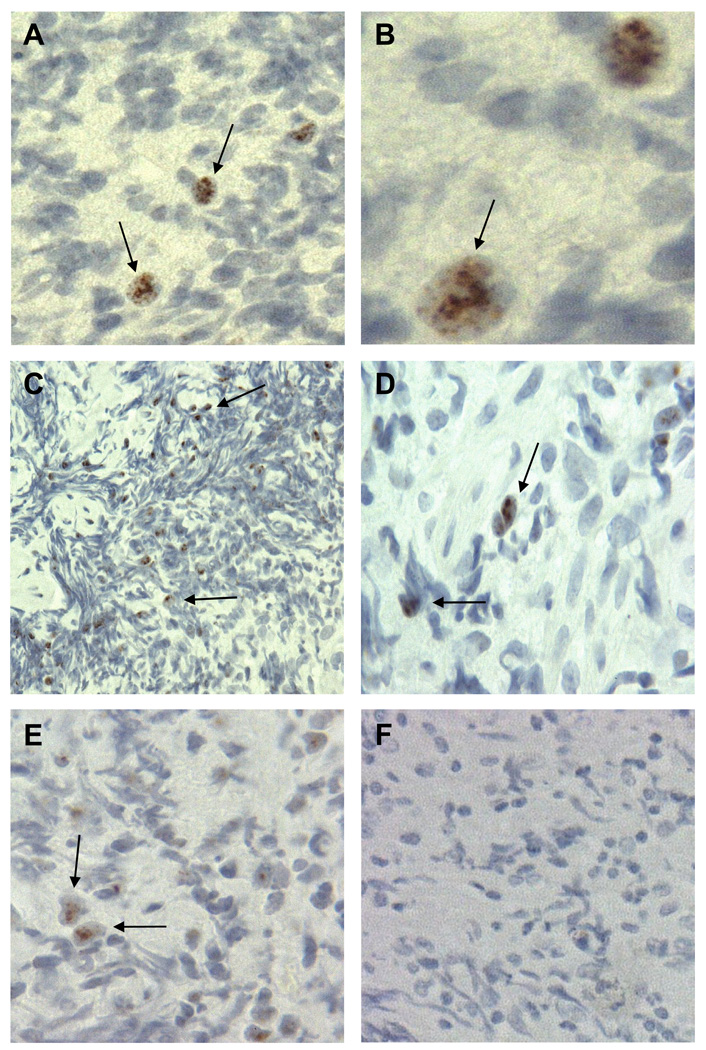
Fig. 1.
SV40 T-ag expression in NHL. (A, B) Expression of SV40 T-ag in an SV40 DNA-positive diffuse large B-cell lymphoma from a 48-year-old white male. (C, D) Expression of SV40 T-ag in an SV40 DNA-positive B-cell lymphoma from a 47-year-old Hispanic male. (E) Expression of SV40 T-ag in an SV40 DNA-positive diffuse large B-cell lymphoma from a 23-year-old Hispanic male. (F) No T-ag expression in an SV40 DNA-negative B-cell lymphoma from a 39-year-old Hispanic male. Samples were stained with antibody PAb101. Arrows point to representative SV40 T-ag-positive cells in panels A–E. Original magnification for panels A, C, E, and F, 40×, and for panels B and D, 100×.

