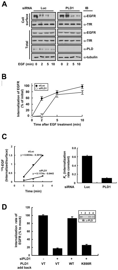
Figure 1. Wild type but not lipase inactive PLD1 facilitates EGFR endocytosis.
(A) HeLa cells were transfected with control (Luc) or PLD1 (PLD1) siRNA to deplete endogenously expressed PLD1. After serum starvation for 12 h, EGF was treated at 20 nM for 0, 2, 5, or 10 min and biotinylation of cell surface proteins was performed. Biotinylated (Cell surface) proteins were separated using streptavidin beads and analyzed by western blotting. A representative immunoblot of three independent experiments is shown. (B) Quantitation of EGFR internalization in HeLa cells. The kinetics of the EGF-induced internalization of EGFR in HeLa cells transfected with either luciferase (closed circle) or PLD1 (open circle) siRNA. (C) HeLa cells depleted of endogenous PLD1 were incubated with 125I-EGF at 37°C. ke valueswere measured during linear 3-min time course and expressed as per cent of the mean values obtained for control-transfected cells. The data represent mean values from three independent experiments and the error bars represent standard deviations. (D) After depleting endogenous PLD1 using PLD1 siRNA, endogenous level of wild type or K898R PLD1 was expressed in HeLa cells. The internalization rate of EGFR was calculated as described in (C) and is shown as a bar graph reflecting the average of three independent experiments and standard deviations. Immunoblots in the inset indicate the expression levels of the constructs used.

