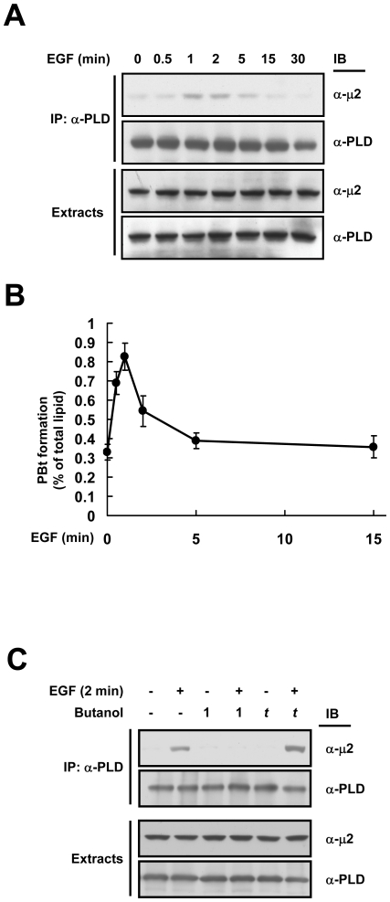
Figure 2. PLD1 associates with the μ2 subunit of AP2 in a PA-dependent manner.
(A) HeLa cells were treated with EGF (20 nM) and cell extracts were immunoprecipitated with anti-PLD antibody and then immunoblotted with the indicated antibodies. (B) The kinetic of EGF-induced PLD1 activation in HeLa cells. (C) HeLa cells were treated with EGF (20 nM) for 1 min in the absence or presence of either 1-butanol (0.4%) or t-butanol (0.4%) as shown and then the cell lysates were immunoprecipitated as in (A). Alcohols were added 5 min prior to EGF treatment.

