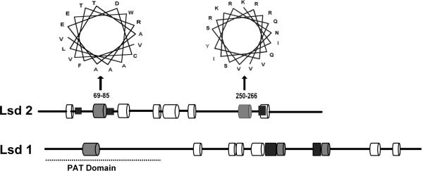
Figure 3. Predicted α-helical regions of Lsd2.
The locations of the eight predicted α-helices of Lsd2 (352aa) are indicated with cylinders: 36–44, 69–85, 110–114, 143–152, 153–176, 187–197, 250–266 and 275–286. Grey cylinders represent putative lipid-binding helices. Black rectangles represent hydrophobic segments that could also interact with the lipid droplet. Helical wheel diagrams of the two predicted lipid binding helices are shown. A scheme of the helices found in Lsd1 is shown for comparison.

