
Figure 4. Amino acid sequence alignment of Lsd1 and Lsd2 from Drosophila melanogaster.
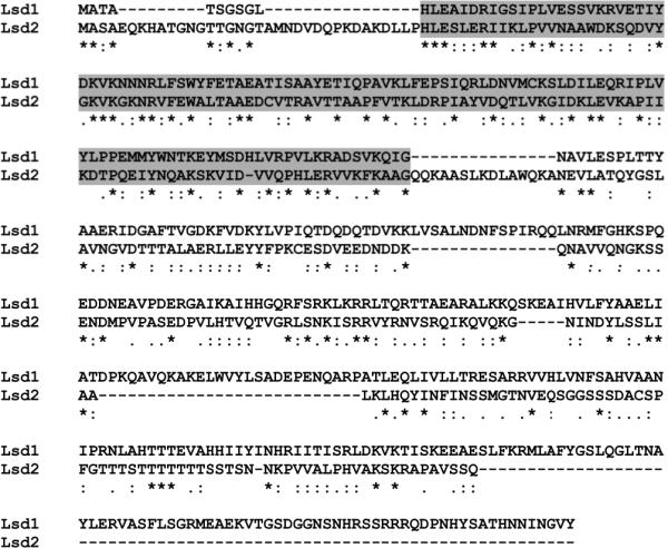
The alignment was performed using the T Coffee program available at www.ebi.ac.uk/t-coffee. Lines in the sequence are gaps introduced by the program to optimize the alignment. Identical residues in all the sequences are denoted by asterisks; conservative substitutions are denoted by dots. Conserved PAT domains are boxed in gray.