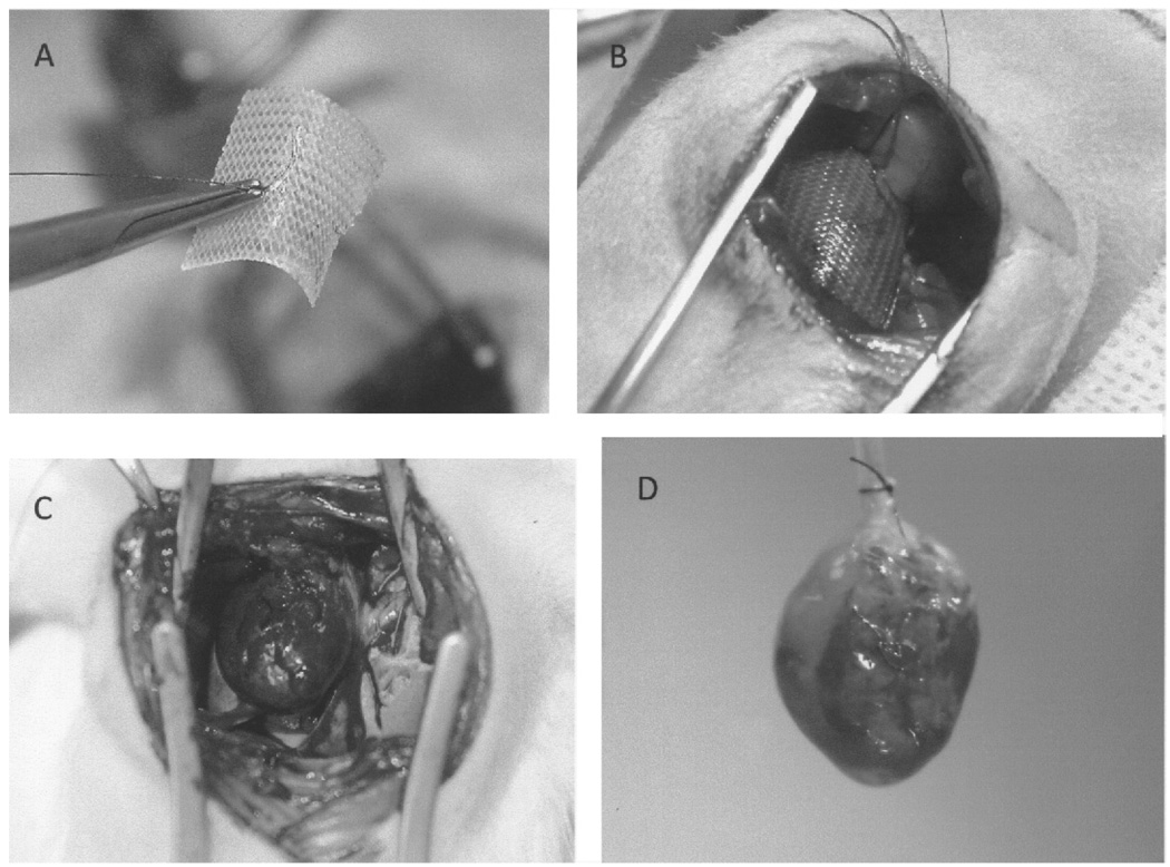
Figure 2.
(A) Three-dimensional fibroblast culture (3DFC) prior to implantation; the suture in the middle of the patch is used to attach the 3DFC to the left ventricle. (B) 3DFC at the time of implantation on the infarcted left ventricle. (C) 3DFC at 3 weeks after myocardial infarction. Note that the 3DFC is well integrated and attached to the infarcted wall. (D) 3DFC in a perfused heart preparation at 3 weeks after myocardial infarction. As note above, the 3DFC is well integrated into the infarcted wall and the suture is easily visible.

