
Figure 1.
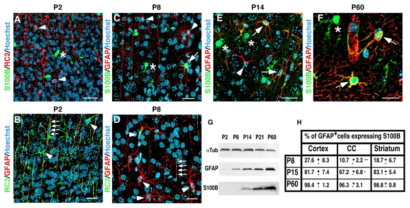
S100B is expressed long after the gain of GFAP and the loss of RC2 and characterizes a mature developmental stage of the astrocytic lineage. A–E, coronal sections of developing post-natal telencephalon of mice at P2, P8, P14 and P60 were subjected to double immunolabeling as indicated. A–B, at P2, RC2+ transforming RGCs do not yet express S100B (A, arrowheads) but start to express GFAP in their radial processes (B, arrows). In B, arrowheads point to RC2+ RGC bodies which are still negative for GFAP staining. C–D, at P7, RG transforming cells are more stellate and express GFAP in their cell bodies (C and D, arrowheads). Some of these cells start to express S100B (C, arrow). In D, arrows point to a radial process with residual RC2 staining. E, at P14, most of GFAP expressing cells express S100B (arrows). The arrow points to a GFAP+ cell that does not yet express S100B. F, at P60, all GFAP+ cells express S100B (arrows). In A, C, E and F, asterisks indicate S100B expressing cells that do not express RC2 or GFAP. These cells likely are oligodendroglial cells as indicated in supplemental figure 1. G, Western blot analysis of GFAP and S100B expression during the forebrain postnatal development. α-tubulin (αTub) was used as loading control. H, quantitative analysis of GFAP+ cells expressing S100B in different telencephal regions including the cortex, the corpus callosum (CC) and the striatum during postnatal development. 3 mice were analyzed at P8 and P14 and 5 at P60. Errors are ± SD. ** p<0.001, * p<0,05. Scale bars: 20 μm.