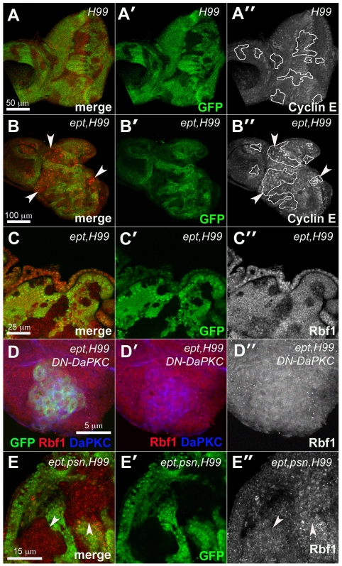
Figure 3. ept mutations reduce levels of the Drosophila Rb ortholog Rbf1.
Confocal images of larval eye discs containing clones of H99 mutant cells (A–A″), ept,H99 double-mutant cells (B–C″) or ept,H99,psn triple mutant cells (E–E″) marked by the absence of GFP (green) co-stained for CycE (red in A–B″), or Rbf1 (red in C–C″, and E–E″). Tracing in A″ and B″ outlines H99 and eptH99 mutant clones respectively. The anti-CycE signal was recorded at the same optical settings in panels A″ and B″. Arrowheads in panels B and B″ denote ept,H99 double mutant clones that express CycE. (D–D″) MARCM-mediated expression of DN-DaPKC (blue) in ept,H99 double-mutant cells marked by GFP (green) restores levels of Rbf1 (red). Arrows in panel E denote clones of ept,H99,psn cells that express normal levels of Rbf1 relative to adjacent control cells.

