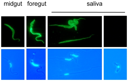
Figure 3. Expression of PSSA-2 in tsetse-derived trypanosomes.
Immunofluorescence analysis of PSSA-2-HA expression by trypanosomes isolated from the midgut, foregut and saliva probes 28–35 days after the infective bloodmeal. Trypanosomes were fixed with formaldehyde and glutaraldehyde and stained with anti-HA antibody (upper panel) and DAPI (lower panel).

