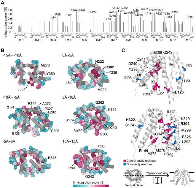
Figure 3. High-IS residues of LacY.
(A) IS pattern of LacY. Black line corresponds to the 90th percentile of IS. Transmembrane regions are indicated as helices below the x-axis with boundary residue numbers; 25 detected residues are labeled with residue numbers. (B) Serial sections of LacY structure from cytoplasm (−15Å) to periplasm (15Å). The detected residues are shown as vdW spheres with residue numbers; 5 irreplaceable residues are shown in bold. (C) ‘Open book’ view of the detected residues in LacY. Central cavity and non-cavity residues are shown in red and blue sticks, respectively; five irreplaceable residues are indicated as bold characters. Transmembrane helix numbers are shown in roman numerals.

