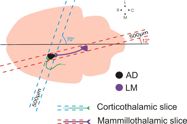
Figure 1.
Schematic illustration of the blocking angles used to generate the two slice preparations. The mammillothalamic slice was prepared as follows: taking the midline as a reference, a 12° cut was made to parallel the route of the mammillothalamic tract and a single 500-μm-thick slice was vibratomed. The slice containing the severed cortical axons into AD was prepared by blocking the brain at a 70° angle and by cutting a single 500-μm-thick slice. C, Caudal; L, lateral; M, medial; R, rostral.

