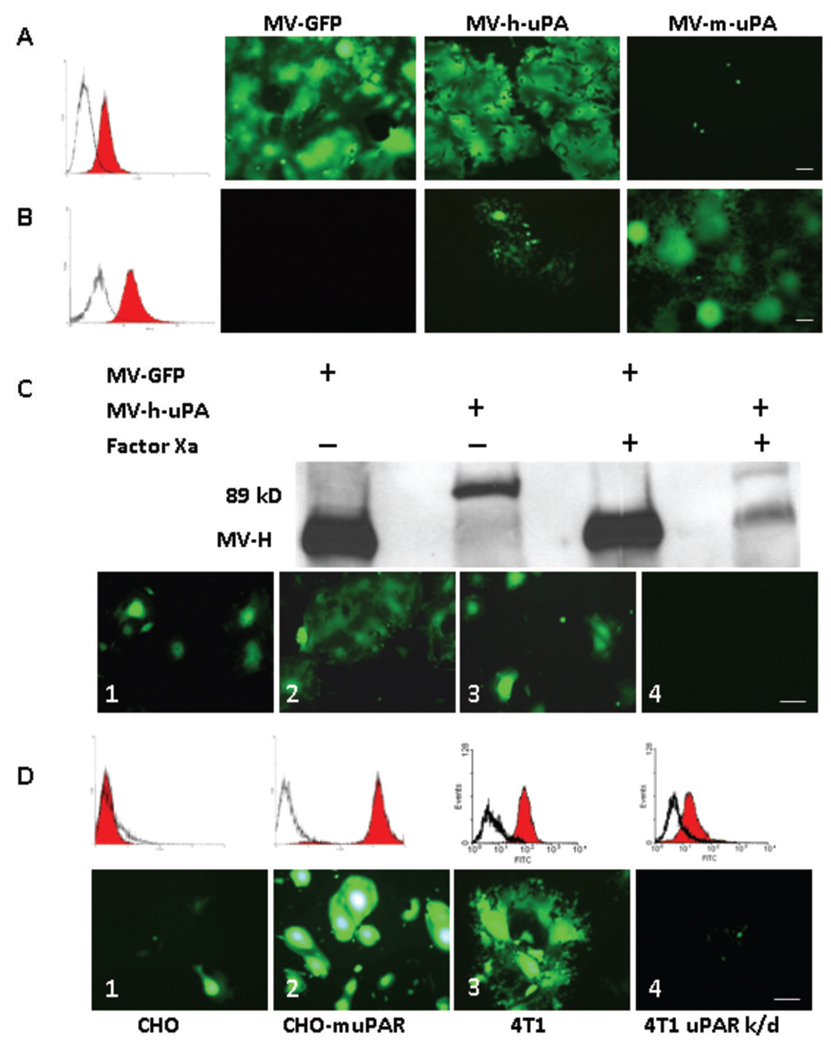
Figure 2. Receptor and species specificity of MV-uPA.
uPAR expression in human MDA-MB-231 (A) and MC-38 (B) was assessed with human and murine anti-uPAR monoclonal antibodies (filled histograms) or isotype controls (open histograms). Cells were infected with each of recombinant measles viruses as indicated at an MOI of 0.5 and photographed 48 h after infection. MDA-MB-231 cells (A) underwent cell fusion after MV-GFP and MV-h-uPA infection, but not with MV-m-uPA. Conversely, MC-38 cells (B) were resistant to unmodified MV-GFP and MV-h-uPA (isolated green cells but not significant syncytia were observed-due to some degree of cross reactivity between human and mouse uPAR) and sensitive to MV-m-uPA. C. The chimeric MV-H-ATF glycoproteins contain a (coagulation) factor X(a) cleavage linker (IEGR) (32), before the NotI restriction site (Fig. 1. A). An aliquot of MV-GFP or MV-h-uPA viral particles was pretreated with 20 µg/mL of activated factor X [FX(a)] (New England Biolabs, Beverly, MA) or PBS (mock treatment) for 30 minutes at room temperature, in sterile conditions, before infection of MDA-MB-231 cells. Western blot analysis shows successful factor X(a) induced cleavage of the linker and detachment of the uPA-ATF from the H glycoprotein (western blot, far right lane). Untreated MV-GFP and MV-h-uPA (MOI= 0.5) induced cell fusion in MDA-MB-231(Lower panel, C.1, MV-GFP; C.2, MV-h-uPA). Factor X(a) treatment of MV-GFP did not affect cell fusion (C. 3). Factor X(a) treatment of MV-h-uPA prevented fusion and syncytia formation in MDA-MB-231 cells (C.4). D. Chinese hamster ovary (CHO) cells stably overexpressing murine uPAR (D.2) underwent fusion and syncytia formation after infection with MV-m-uPA, compared to wild type CHO cells (D.1). D.3. 4T1 cells express murine uPAR and undergo fusion after infection with MV-m-uPA (MOI=1). uPAR expression in this cell line was knocked down by a retroviral vector encoding microRNA-based shRNA against mouse uPAR. uPAR expression was significantly decreased as determined by real time PCR (data not shown) and by flow cytometry (86% decrease, as assessed by FACS analysis of uPAR). uPAR knockdown (D.4) of 4T1 cells reduces the ability of MV-m-uPA to induce cell fusion. Scale bar = 100 µm.

