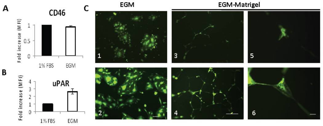
Figure 3. uPAR dependent in vitro endothelial cell infection.
HUVECs were grown in full endothelial growth medium (EGM-2) (stimulated) or in EBM-2 medium with 1% FBS (unstimulated). Stimulation of HUVEC monolayers with 2% serum and growth factors (endothelial growth medium, EGM-2) was associated with upregulation of uPAR (B), but not CD 46 (A), compared to unstimulated HUVECs (endothelial basal medium and 1% FBS). Changes in HUVEC expression of CD46 and uPAR were determined by FACS analysis, and displayed as fold increase of mean fluorescence index (MFI) before and after stimulation. HUVEC monolayers were infected with viruses at an MOI=0.5. C. In stimulated HUVECs, MV-h-uPA induced cell fusion more efficiently than MV-GFP (C. 2, vs. C. 1). Scale bar = 100 µm. HUVECs (grown in EGM-2) were plated on matrigel and tubes were allowed to form (16 hours). Once tubes were formed, they were infected with either MV-GFP (C. 3, C.5) or MV-h-uPA (C. 4, C. 6) at a MOI = 1. Pictures of the areas of abundant tubes mostly in the center of wells were taken at 72 hours after infection. MV-h-uPA was associated with more efficient capillary infection compared with the unmodified virus control (C. 4 and 6 vs. C. 3 and 5). Experiments were done in triplicate. Scale bar = 500 µm (C. 3, 4) and 50 µm (C. 5, 6).

