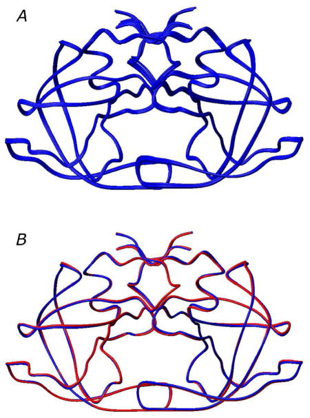
Figure 4.
Lowest energy HIV-1 protease dimer structures. (A) Best-fit superposition of the 10 lowest energy structures from the cluster containing the lowest energy structure. (B) Comparison of the restrained regularized mean structure (blue), derived from the 10 lowest energy structures, with the subunit arrangement in the X-ray structure51 (red). ( .)

