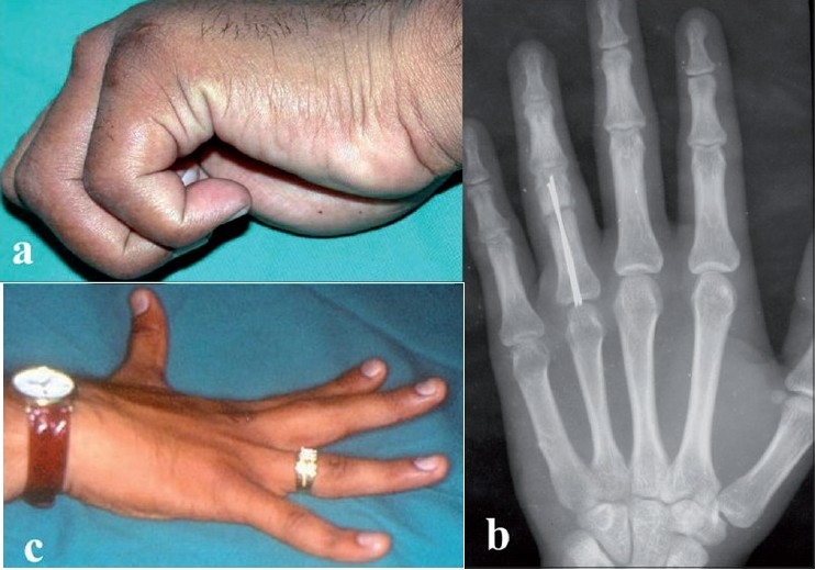
Figure 5.

(a) Clinical photograph of adventitious bursitis at the site of entry portal in one patient (b) X-ray depecting of backing of nails causing adventitious bursitis. (c) Clinical photograph of 5° extension lag at proximal interphalangeal joint in one of the patients
