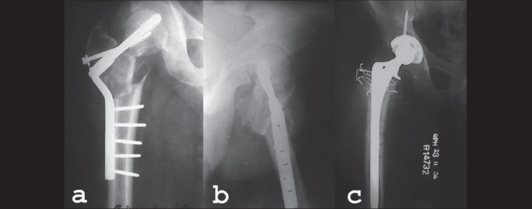
Figure 1.

(a) AP radiographs of 38yrs old gentleman. Fracture neck of femur Rt.side treated with primary Powell's osteotomy and fixation with double angle barrel plate. (b) Lateral radiograph of same patient showing implants cutting out and fracture fragments in malaligned position. (c) AP radiograph showing uncemented total hip arthroplasty done 14 months after the primary fixation surgery
