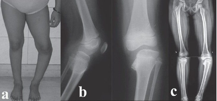
Figure 1.

(a) Preoperative clinical photograph of an eight-year-old girl showing a varus deformity of the left proximal tibia. (b) Preoperative lateral and anteroposterior radiographs showing medial beaking and a significant depression of the medial tibial epiphysis and metaphysis (Langenskiold Stage IV). (c) Preoperative alignment-view radiograph of both lower limbs showing a medial mechanical axis deviation and demonstrating the extent of the deformity
