Figure 4.

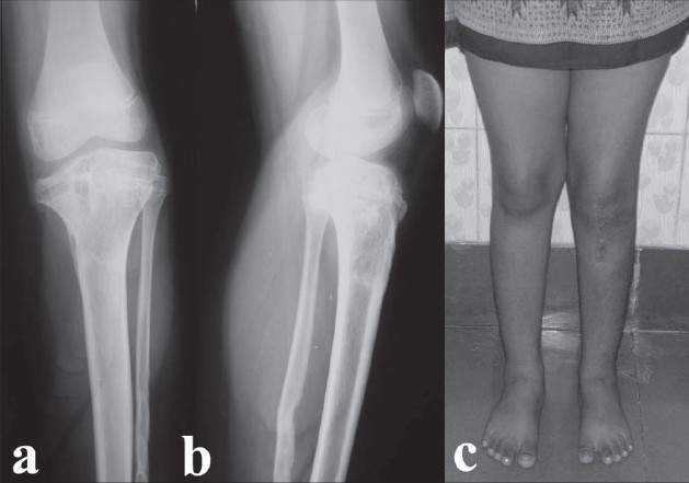
Radiographs (a,b) at final follow-up after percutaneous proximal tibial and fibular epiphysiodesis. Clinical photograph (c) at final (four-year) follow-up showing full correction of deformity and excellent alignment

Radiographs (a,b) at final follow-up after percutaneous proximal tibial and fibular epiphysiodesis. Clinical photograph (c) at final (four-year) follow-up showing full correction of deformity and excellent alignment