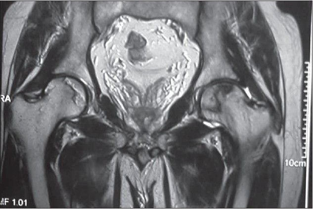
Figure 1B.

T2 weighted coronal section shows avascular necrosis involving bilateral femoral heads with reparative border appearing hypo/hyperintense (double rim sign) on right side indicating granulation and sclerosis respectively. Dark line bordering the AVN focus on left side represents sclerosis. The cartilage and acetabulum are normal.
