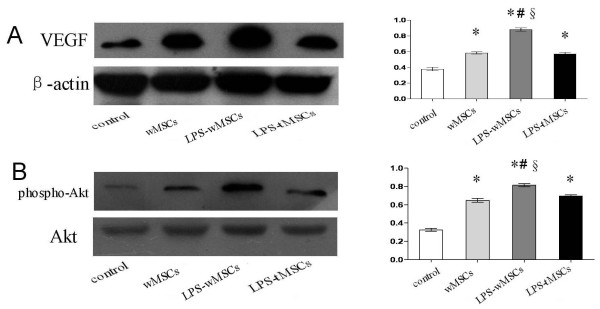
Figure 6.
LPS-preconditioned MSCs transplantation increased expression of VEGF and phosphorylated Akt protein in vivo. Representative Western blots for expression of VEGF. Corresponding β-actin blots are shown as a control for sample loading. Bar graph on right shows quantitation. The protein levels for each sample were determined as a ratio to their corresponding β-actin levels. B. Representative Western blots for expression of phospho-Akt, Akt. Total Akt served as a loading control. Bar graph on right shows quantitation. The protein levels of phospho-Akt for each sample were determined as a ratio to their corresponding Akt levels. Data are mean ± SEM. *P < 0.05 versus control group; #P < 0.05 versus wMSCs group; §P < 0.05 versus LPS-tMSCs group.

