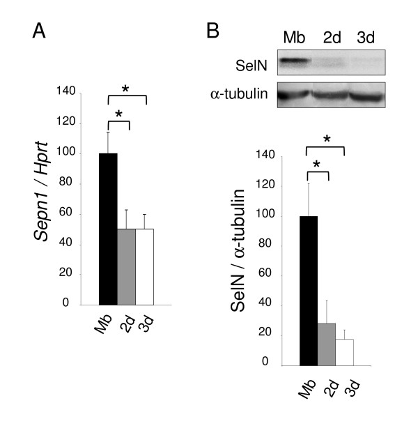
Figure 3.
Sepn1 expression during murine myoblast differentiation. A: Sepn1 qRT-PCR on cDNA from C2C12 myoblasts (Mb) and myotubes after 2 and 3 days (2 d/3 d) in differentiation medium. Hprt served for normalization. A two fold reduction of Sepn1 expression was observed in myotubes compared to undifferentiated cells. B: SelN Western blot analysis on proteins from C2C12 cells (Mb, 2 d, and 3 d), normalized to α-tubulin. Note that the decrease of SelN expression between myoblasts and myotubes is even more marked compared to transcript quantification. *, p < 0.05.

