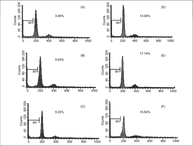
Figure 7.
Flow cytometric analysis of DNA content for detection of apoptosis after 24 and 72 h of melatonin treatment. The cells were plated at 10 × 105 cells/flask and cultured in osteogenic medium in presence of melatonin. A, D after 24, 72 h treatment with osteogenic medium alone, respectively. B, E: after 24, 72 h treatment with osteogenic medium in presence of melatonin 0.01 nM, respectively. C, F: after 24, 72 h treatment with osteogenic medium in presence of melatonin 10 nM, respectively

