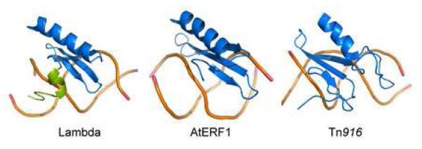
Figure 4. Comparison of three-stranded beta-sheet DNA binding proteins.
The image compares the structures of the DNA complexes of the following proteins: IntN from bacteriophage lambda (left), the ethylene responsive factor domain 1 from Arabidopsis thaliana (AtERF1) 22 and the DNA binding domain from the integrase protein encoded by the Tn916 transposon (IntTn916) 20; 21. Only the backbone of the DNA is shown. Each protein is colored blue, with the exception of the amino-terminal tail of IntN that becomes ordered upon DNA binding, which is colored green. None of the proteins share significant primary sequence homology with one another.

