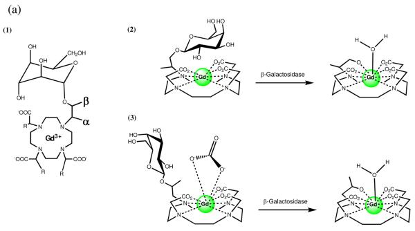
Figure 2.
(a) Schematic representations of the two isomers of enzyme-responsive MR agents activated by β-galactosidase: (1) Flat projection with α and β positions labeled (2) α-EGadMe and (3) β-EGadMe. Each isomer provides an increase in relaxivity following cleavage of the sugar by different mechanisms. (b) MRI detection of β-galactosidase mRNA expression in living X. laevis embryos. MR images of two embryos injected with EgadMe at the two-cell stage. (A) Unenhanced MR image. The embryo on the right was injected with β-gal mRNA, resulting in the higher intensity regions. The signal strength is 45–65% greater in the embryo on the right containing β-gal (contrast-to-noise ratio ranges from 3.5 to 6). The cement gland has intrinsically short T1, thus is visible as a bright structure on both embryos. (B) Pseudocolor rendering of same image in (A) with water made transparent. The image correction makes it possible to recognize the eye, and brachial arches in the injected embryo: d, dorsal; v, ventral; r, rostral; e, eye; c, cement gland; s, somite; b, brachial arches. Scale bar = 1 mm.


