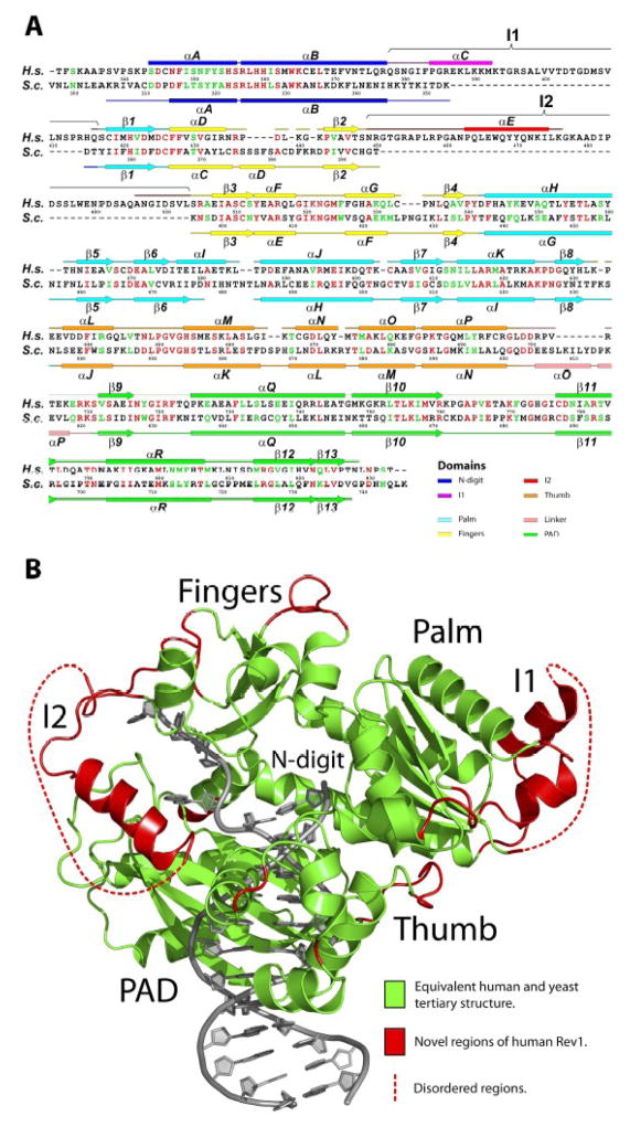
Fig. 2.
Comparison of human and yeast Rev1. (A) Sequence and secondary structure comparison between the catalytic cores of human (H.s) and yeast (S.c) Rev1. Identical residues are highlighted in red and semi-conserved residues are shown in green. (B) Human Rev1 catalytic core displayed in two colors: green, for elements that are conserved between the human and yeast Rev1; red, for elements (including, inserts I1 and I2) that are unique to human Rev1. The DNA is shown in gray.

