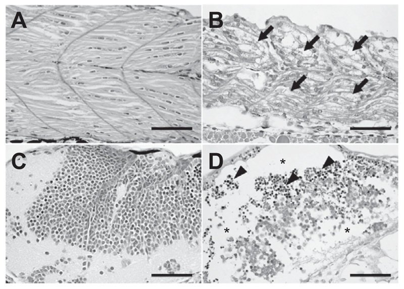
Figure 4.

Histopathological examination. The zebrafish embryos at 24 h post-fertilization were exposed to genistein or vehicle for 60 h. The zebrafish embryos that survived after 0.25 × 10⁻⁴ M genistein or vehicle treatments were prepared for histopathological examination. Histopathological examination revealed that (arrows) granular degeneration of (B) myocytes in skeletal muscle in addition to (asterisks) loss and (arrow heads) apoptosis of (D) neural cells in the brain of the 0.25 × 10⁻⁴ M genistein-treated group. (A and C) vehicle-treated zebrafish embryos, (B and D) genistein-treated zebrafish embryos. Bar = 50 μm.
