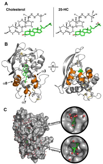
Figure 1. Structure of NPC1(NTD) Bound to 25-Hydroxycholesterol.
(A) Stick models of cholesterol (left) and 25-HC (right) with carbon positions numbered. Carbon and oxygen atoms are colored green and red, respectively.
(B) NPC1(NTD) is represented as a ribbon diagram in gray, and the disulfide bonds are shown in yellow. The positions of cholesterol and 25-HC are essentially identical. For simplicity, we show only the 25-HC molecule (colored in green). Helix3, helix7, and helix8 are colored orange.
(C) The surface of NPC1(NTD), colored in gray, reveals openings at either end of the bound sterol. The W-opening would allow passage of a single water molecule (red spheres), but not a sterol molecule. The S-opening would become large enough to allow entry or exit of a sterol if the opening were expanded slightly (see Figure 2D).

