
Fig. 1.
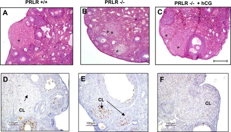
Histological analysis of ovaries from prolactin receptor (PRLR)+/+, PRLR−/−, and human chorionic gonadotropin (hCG)-treated PRLR−/− mice. Representative view of sections of ovaries stained with hematoxylin-eosin from PRLR+/+ (A), PRLR−/− (B), or hCG-treated PRLR−/− (C) of 8-wk-old female mice. Ovaries contain all developmental stages of follicle development. *Corpus luteum (CL). Bar scale: 200 μm. Immunodetection of cleaved caspase-3 in CL of PRLR+/+ (D), PRLR−/− (E), or hCG-treated PRLR−/− (F) mice at 2.5 days postcoitum (dpc). Bar scale 100 μm.