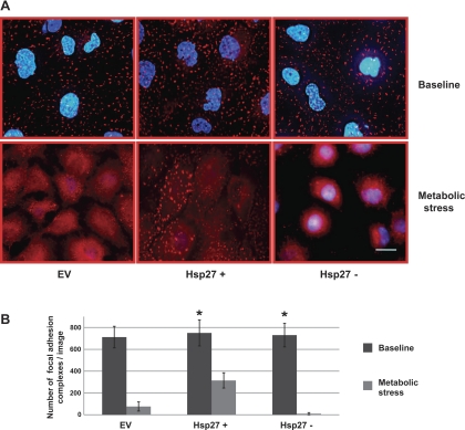
Fig. 5.
Effect of Hsp27 expression on focal adhesion complex integrity during sublethal injury. A: immunocytochemical staining of paxillin (red) assessing the integrity of the focal adhesion complexes at baseline (top) and after metabolic stress (bottom) in EV-treated or Hsp27+ or Hsp27− cells. Nuclei are stained with DAPI (blue). EV- and NSsiRNA-treated cells were not visually distinguishable (NSsiRNA not shown). Images represent random fields from 3 independently repeated experiments. Scale bar = 5 μm. B: number of intact focal adhesion complexes per field counted with Image J. Minimum particle size: 50 pixels2. Data are means ± SE and represent 5 random fields from 3 separate experiments; (n = 15). *P < 0.05 vs. EV.

