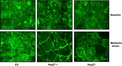
Fig. 6.
Effect of Hsp27 expression on the actin cytoskeleton during sublethal injury. Immunofluorescence micrographs of green fluorescent phalloidin staining of F-actin filaments at baseline (top) and after metabolic stress (bottom) in EV-treated or Hsp27+ or Hsp27− cells are shown. After stress, disruption of the F-actin cytoskeleton and formation of rhodamine-positive, actin-containing aggregates are evident. EV- and NSsiRNA-treated cells were not visually distinguishable (NSsiRNA not shown). Images represent random fields from 3 independently repeated experiments. Scale bar = 5 μm.

