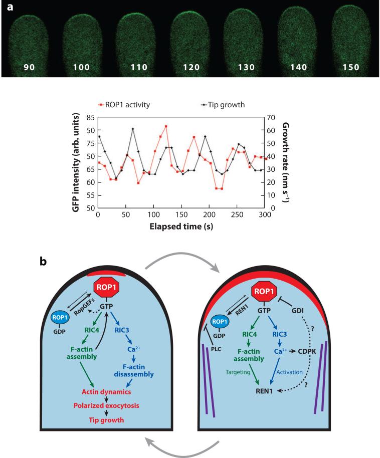
Figure 4.
A model for the generation and maintenance of the dynamic and oscillating apical cap of active ROP1 (where ROP refers to Rho-related GTPase from plants) in pollen tubes. (a) A time series of images showing the localization of a green fluorescent protein (GFP)-based active ROP1 reporter in a tobacco pollen tube transiently expressing this marker, i.e., GFP fused to the N terminus of a RIC4 deletion mutant (Hwang et al. 2005). Numbers at the bottom of each pollen tube image indicate seconds in the time series shown in the graph at the bottom of this figure. (Bottom) Graphs show the oscillation of plasma membrane (PM)-localized GFP-RIC4ΔC. Modified from figure 3 of Hwang et al. (2005). (b) Models for positive feedback–mediated lateral propagation of the apical ROP1 activity and for negative feedback–mediated global ROP1 downregulation by the REN1 GTPase–activating protein (RhoGAP) and lateral inhibition by cortical microtubules (MT) and phospholipase C (PLC). Solid arrows indicate pathways supported by experimental evidence. Dotted arrows indicate speculative pathways lacking experimental support. Abbreviations used: CDPK, calcium-dependent protein kinase; F-actin, actin microfilaments; RIC, ROP-interactive Cdc42/Rac-interactive binding (CRIB) motif–containing protein.

