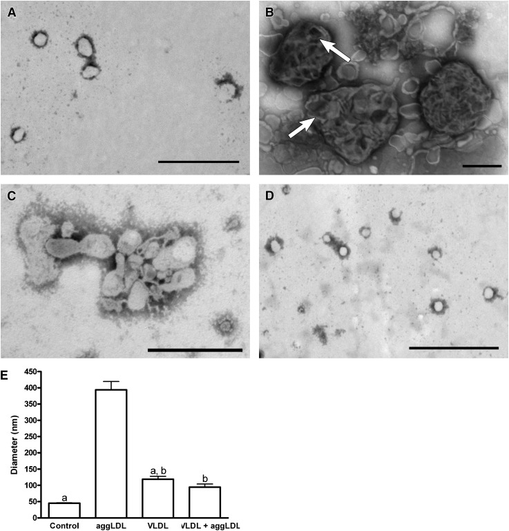
Fig. 4.
THP-1 macrophages treated with TRPs for 6 days had reduced lysosome diameter compared with macrophages that were only cholesterol enriched. A–D: Negative stain electron micrograph of lysosomes isolated from control cells (A), aggLDL-treated cells (100 μg aggLDL protein/ml; B), VLDL-treated cells (100 μg VLDL protein/ml; C), or cells coincubated with aggLDL and VLDL (100 μg aggLDL protein/ml and 100 μg VLDL protein/ml; D). E: The average lysosomal diameter determined from three separate experiments. Lysosomes from control cells were generally small and had a homogenous appearing lumen (A and E). In contrast, lysosomes from aggLDL-treated cells had significantly larger (P < 0.05) lysosomes that had a variety of appearances but often contained a heterogenous mixture of apparently undigested material (arrows in B) within their lumen (B and E). Lysosomes from VLDL-treated cells were small with homogenous lumens similar to those isolated from control cells (C and E). Significantly, when THP-1 were incubated with VLDL in combination with aggLDL, their lysosomes remained small and failed to develop the large, heterogenous appearance of lysosomes isolated from cells incubated with aggLDL alone. Within each panel, bars with the same letter indicate that means were not statistically different. All other comparisons were significantly different (P < 0.05). Magnification for A, C, and D = 40,000×; magnification for B = 25,000×; bar = 500 nm.

