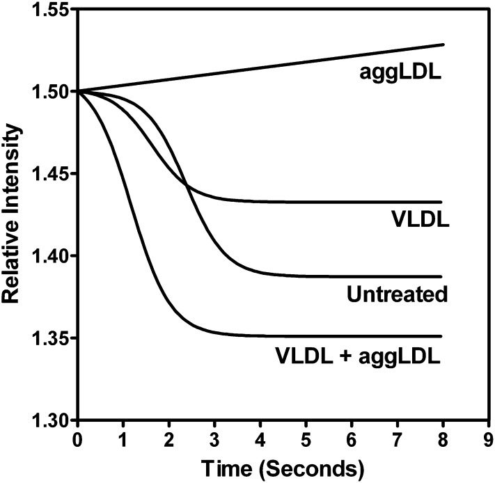
Fig. 7.
Activation of v-ATPases in isolated lysosomes following TG and/or cholesterol enrichment. Untreated lysosomes exhibited activation of v-ATPase and the pumping of hydrogen ions into the lysosomal lumen when stimulated by the addition of ATP and valinomycin (time 0), as indicated by the decrease in the relative fluorescence intensity of acridine orange. Lysosomes from macrophages that had been treated with 100 μg aggLDL protein/ml exhibited a lack of v-ATPase activity as evidenced by no reduction in the relative fluorescence intensity. In contrast, lysosomes from cells treated with 100 μg VLDL protein/ml, either alone or in combination with 100 μg aggLDL protein/ml, exhibited rapid quenching of acridine orange fluorescence, indicating active v-ATPases. Data are a representative of example chosen from multiple separate experiments.

