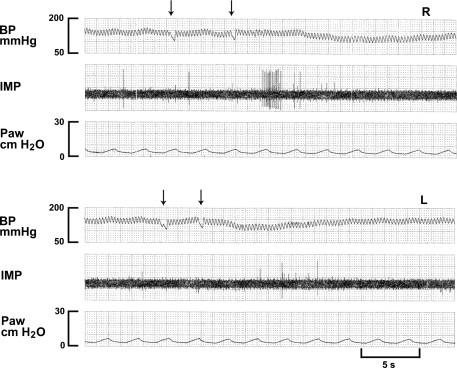
Fig. 3.
A C-fiber receptor (CFR) in response to injection of lidocaine (4 mg/kg) into the right ventricle (top 3 traces, R) and left ventricle (bottom 3 traces, L). Note that this CFR has lower background activity (almost inactive). In contrast to mechanosensors, the CFR is stimulated instead of inhibited following injection of lidocaine (arrows denoting start and end of the injection) into the pulmonary circulation.

