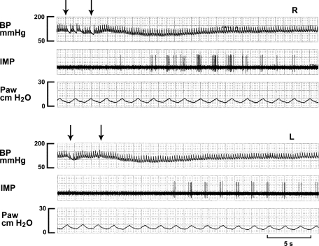
Fig. 4.
A high-threshold Aδ-receptor (HTAR) in response to injection of lidocaine (4 mg/kg) into the right ventricle (top 3 traces, R) and left ventricle (bottom 3 traces, L). Note that this HTAR has lower background activity. It is stimulated by both injections into the right and left ventricles (arrows denoting injection period) with a longer latency for left-side injection; this indicates a preferential perfusion by pulmonary circulation.

